Creating an Animal Cell Coloring Worksheet

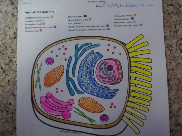
Animal cell coloring biology corner – Designing an effective animal cell coloring worksheet requires careful consideration of both visual appeal and educational value. A well-designed worksheet should accurately represent the cell’s structure while engaging students through color and visual cues, facilitating a deeper understanding of organelle function and location.
The worksheet should present a simplified, yet accurate, representation of an animal cell. Overly complex diagrams can be overwhelming for younger learners, while simplistic drawings may lack sufficient detail for older students. Finding a balance between detail and simplicity is crucial for effective learning. The use of color-coding and visual textures enhances comprehension and memorization.
Organelle Representation and Descriptions
The worksheet should feature a large, central cell Artikel. Within this Artikel, each major organelle should be clearly depicted and labeled. Organelles should be drawn in a size proportional to their relative size within the cell, though artistic license can be used to enhance clarity. Each organelle should be assigned a unique color, and this color-coding should be consistent throughout the worksheet.
For example, the nucleus could be depicted as a large, round, purple structure with a slightly textured appearance to represent the chromatin. The description alongside it would read: ” Nucleus (Purple): The control center of the cell, containing the cell’s genetic material (DNA).” Similarly, the rough endoplasmic reticulum could be represented as a network of interconnected, slightly bumpy, blue lines to visually depict the ribosomes attached to its surface.
Its description might be: ” Rough Endoplasmic Reticulum (Blue): A network of membranes studded with ribosomes; involved in protein synthesis and transport.”
Okay, so we’re totally into this animal cell coloring thing from Biology Corner, right? It’s like, seriously detailed. But sometimes you need a break from the organelles and whatnot. Check out these awesome animal coloring sheets for kids for a fun, simpler approach before diving back into the complexities of the cell membrane and cytoplasm. It’s a good way to refresh before tackling those tricky Golgi apparatus diagrams back in Biology Corner.
Color Key and Visual Cues
A clearly defined color key is essential. This key should list each organelle, its corresponding color on the worksheet, and a brief description. The color key could be presented as a table for easy reference. For instance:
| Organelle | Color | Description |
|---|---|---|
| Nucleus | Purple | Control center; contains DNA |
| Rough Endoplasmic Reticulum | Blue | Protein synthesis and transport |
| Smooth Endoplasmic Reticulum | Light Blue | Lipid synthesis and detoxification |
| Golgi Apparatus | Yellow | Packages and modifies proteins |
| Mitochondria | Red | Powerhouse of the cell; produces ATP |
| Lysosomes | Green | Waste disposal and recycling |
| Ribosomes | Dark Blue (dots on RER) | Protein synthesis |
| Cell Membrane | Brown | Outer boundary; controls what enters and exits |
| Cytoplasm | Light Tan | Gel-like substance filling the cell |
Incorporating visual cues beyond color enhances understanding. For example, the rough endoplasmic reticulum’s bumpy texture visually represents the ribosomes attached to it. The Golgi apparatus could be depicted as a series of flattened sacs, reflecting its actual structure. Using different line weights or patterns can further differentiate organelles and improve visual clarity. The mitochondria’s bean-shaped structure should be accurately depicted, helping students visualize its morphology.
Illustrating Animal Cell Components: Animal Cell Coloring Biology Corner

Accurate depiction of animal cell components is crucial for understanding their functions and interactions. A well-illustrated diagram should clearly show the relative sizes and locations of organelles within the cell, emphasizing their structural features and relationships. This section details the visual representation of key animal cell components.
The Nucleus
The nucleus, the cell’s control center, should be illustrated as a large, roughly spherical organelle. Its prominent feature is the nuclear envelope, a double membrane punctuated by nuclear pores. These pores, though small, should be visible, indicating the selective transport of molecules between the nucleus and cytoplasm. The nucleolus, a denser region within the nucleus, should be shown as a smaller, round structure.
It is the site of ribosome biogenesis and should be depicted as a distinct area within the nucleus. The overall appearance should convey the nucleus’s importance as the repository of genetic material and the regulator of cellular activities.
Mitochondria
Mitochondria, the “powerhouses” of the cell, are depicted as elongated, bean-shaped organelles with a characteristic double membrane structure. The outer membrane should be smooth, while the inner membrane should be illustrated with numerous folds called cristae. These cristae significantly increase the surface area for cellular respiration. The matrix, the space within the inner membrane, should be shown as a relatively homogenous area.
The illustration should clearly show the double membrane structure to highlight the compartmentalization crucial for ATP production. The number of mitochondria shown should reflect their abundance in actively metabolizing cells.
Ribosomes, Animal cell coloring biology corner
Ribosomes are depicted as small, dark dots scattered throughout the cytoplasm and also attached to the endoplasmic reticulum. Their small size should be contrasted with the larger organelles. The illustration should clearly distinguish free ribosomes (in the cytoplasm) from those bound to the endoplasmic reticulum. Their function in protein synthesis should be emphasized by their proximity to the endoplasmic reticulum and the Golgi apparatus, which are involved in protein processing and transport.
The high density of ribosomes in areas of active protein synthesis should be reflected in the illustration.
The Endoplasmic Reticulum and Golgi Apparatus
The endoplasmic reticulum (ER) should be illustrated as a network of interconnected membranous sacs and tubules extending throughout the cytoplasm. The rough ER, studded with ribosomes, should be distinguished from the smooth ER, which lacks ribosomes. The smooth ER should appear as a network of tubules, while the rough ER should appear as flattened sacs with ribosomes attached. The Golgi apparatus, closely associated with the ER, should be depicted as a stack of flattened, membrane-bound sacs (cisternae).
The illustration should clearly show the movement of vesicles, small membrane-bound sacs, budding from the ER and fusing with the Golgi apparatus, illustrating the flow of proteins through these organelles for modification and packaging before transport to their final destinations within or outside the cell. The overall appearance should highlight the interconnectedness of these two organelles in the processing and transport of proteins.
Question & Answer Hub
What are some alternative coloring techniques besides realistic depictions?
Simplified diagrams, cartoonish styles, or even abstract representations can be used, depending on the age group and learning objectives. The goal is to make the activity engaging and accessible.
How can I assess student understanding after a coloring activity?
Use a combination of methods: Observe their coloring choices and accuracy of labeling; ask them to explain the function of different organelles; give a short quiz on cell structure.
Are there any free online resources besides Biology Corner?
Yes, many websites and educational platforms offer free printable worksheets and diagrams related to animal cell structure. A simple online search will reveal numerous options.
What materials are needed besides the worksheet and crayons/colored pencils?
Consider adding supplementary materials like a magnifying glass for observing real-life cell images (if available), or a short video explaining cell function to enhance the learning experience.


