Coloring Keys and Their Purpose: Animal Cell Coloring Key Answers
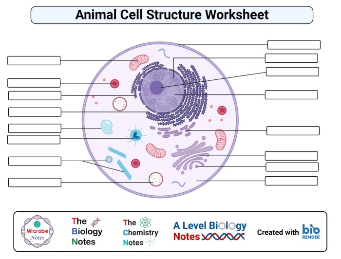
Animal cell coloring key answers – Color plays a crucial role in enhancing the educational value of animal cell diagrams. By assigning different colors to various organelles and structures, students can visually distinguish and understand the complex organization within a cell. This visual learning aid facilitates memorization and comprehension of the different components and their respective functions.Coloring keys provide a guide to the color-coded representations within a diagram.
They ensure clarity and consistency in the visual representation of cellular components, making it easier for students to identify and understand the different parts of an animal cell. A well-designed coloring key acts as a roadmap, connecting the visual representation with the corresponding organelle or structure.
Creating Effective Coloring Keys
A clear and informative coloring key is essential for maximizing the educational benefits of colored animal cell diagrams. Several strategies contribute to creating such a key.
- Distinct Colors: Choose colors that are easily distinguishable from one another to avoid confusion between different organelles. For example, avoid using similar shades of blue for the nucleus and the endoplasmic reticulum.
- Clear Labels: Each color in the key should be clearly labeled with the name of the corresponding organelle or structure. Using both the common name and the scientific name (when appropriate) can enhance understanding.
- Consistent Application: Ensure that the colors used in the diagram consistently match the colors indicated in the key. Any discrepancies can lead to confusion and misinterpretation.
- Logical Grouping: Consider grouping related organelles together in the key. For example, organelles involved in protein synthesis (ribosomes, endoplasmic reticulum, Golgi apparatus) can be grouped together visually in the key, reinforcing their functional connection.
Benefits of Using Coloring Keys
The use of coloring keys offers several benefits in the educational context of animal cell diagrams.
Understanding animal cell structures through coloring diagrams and key answers provides a foundational understanding of biology. While exploring cellular levels, you might also enjoy visually exploring different fauna, such as with these arctic animal coloring pages , offering a creative break. Returning to cell biology, accurate coloring based on the key reinforces learning about organelles and their functions.
- Improved Visual Differentiation: Coloring allows students to quickly and easily differentiate between the various structures within the complex environment of an animal cell.
- Enhanced Memorization: The visual association of colors with specific organelles aids in memory retention and recall of their names and functions.
- Better Understanding of Spatial Relationships: Coloring can highlight the spatial relationships between different organelles, helping students understand how they interact and function together within the cell.
- Engaging Learning Experience: The interactive nature of coloring engages students actively in the learning process, making it more enjoyable and effective.
Common Animal Cell Organelles and Their Colors

Understanding the structure and function of animal cells is fundamental to biology. Color-coding organelles in diagrams or models helps visualize their distinct roles and spatial relationships within the cell. Choosing colors strategically can also aid memorization and enhance understanding.
Organelle Coloring and Rationale
The following table presents suggested colors for common animal cell organelles and the reasoning behind these choices:
| Organelle | Suggested Color | Rationale |
|---|---|---|
| Nucleus | Purple/Dark Blue | Often depicted as the control center, these colors evoke a sense of importance and authority. |
| Mitochondria | Orange/Red | These colors represent energy and activity, reflecting the mitochondria’s role as the “powerhouse” of the cell, generating ATP. |
| Ribosomes | Dark Blue/Brown | These small organelles are responsible for protein synthesis. Darker colors suggest their dense structure and crucial role. |
| Endoplasmic Reticulum (ER) | Light Blue/Pink | These pastel colors represent the ER’s network-like structure involved in protein and lipid synthesis and transport. Different shades can distinguish between rough ER (with ribosomes) and smooth ER. |
| Golgi Apparatus | Yellow/Light Green | These colors depict the Golgi apparatus’s role in processing, packaging, and distributing proteins and lipids. |
| Lysosomes | Green/Brown | These colors suggest the lysosomes’ function in waste breakdown and recycling. |
| Cell Membrane | Light Yellow/Orange | These colors suggest a flexible barrier, representing the cell membrane’s role in regulating what enters and exits the cell. |
| Cytoplasm | Light Gray/Pale Yellow | These neutral colors represent the cytoplasm as the background substance that fills the cell and surrounds the organelles. |
Variations in Animal Cell Structure
While all animal cells share fundamental characteristics, significant variations exist in their structure, reflecting their specialized functions within the organism. This section explores the key differences between animal and plant cells and then delves into the structural variations among different types of animal cells.Understanding the structural variations in animal cells is crucial for comprehending how these cells perform their unique roles, contributing to the overall function of the organism.
These variations are a testament to the adaptability and specialization of cells within the animal kingdom.
Comparison of Plant and Animal Cells, Animal cell coloring key answers
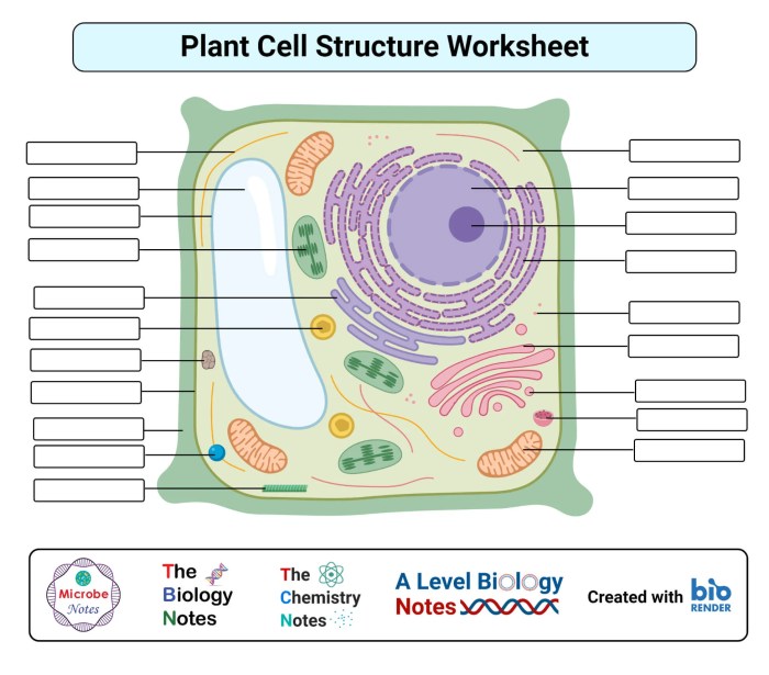
Plant and animal cells, while both eukaryotic, exhibit distinct structural differences. Plant cells possess a rigid cell wall composed primarily of cellulose, providing structural support and protection. Animal cells lack a cell wall but have a flexible cell membrane that allows for changes in shape and facilitates movement. Plant cells also contain chloroplasts, the sites of photosynthesis, which are absent in animal cells.
Furthermore, plant cells typically have a large central vacuole that plays a role in storage and maintaining turgor pressure, while animal cells may have smaller vacuoles or none at all.
Specialized Animal Cell Structures
Different types of animal cells exhibit specialized structures that enable them to carry out their specific functions.Nerve cells, or neurons, are characterized by long, branching extensions called axons and dendrites, which transmit electrical signals throughout the body. These specialized structures facilitate communication between nerve cells and other cells, enabling rapid information processing and coordination of bodily functions.Muscle cells, responsible for movement, contain abundant contractile proteins, such as actin and myosin, organized into filaments.
These filaments slide past each other, causing the muscle cell to shorten and generate force. Variations exist among different types of muscle cells (smooth, skeletal, and cardiac), reflecting their specific roles in different parts of the body.Red blood cells, responsible for oxygen transport, are uniquely shaped biconcave discs lacking a nucleus and most other organelles. This specialized structure maximizes surface area for oxygen binding and allows for efficient passage through narrow capillaries.
Resources for Animal Cell Diagrams and Coloring Keys

Locating accurate and well-labeled animal cell diagrams is crucial for effective learning and teaching. Numerous resources, both online and offline, provide such diagrams and accompanying coloring keys, aiding in the visualization and comprehension of cellular structures and their functions. Selecting appropriate resources requires careful evaluation to ensure accuracy and clarity.
Reliable Sources for Animal Cell Diagrams
Accessing reliable diagrams is essential for accurate understanding. Several reputable sources offer high-quality animal cell diagrams and coloring resources.
- Textbooks: Biology textbooks, particularly those for high school and introductory college courses, are a reliable source. They often include detailed diagrams and explanations of animal cell structure and function.
- Educational Websites: Reputable educational websites like Khan Academy, CK-12, and Biology Corner offer interactive diagrams and activities related to animal cells. These resources often provide clear labeling and explanations.
- Scientific Journals and Publications: Peer-reviewed scientific journals and publications offer the most accurate and up-to-date information on cell biology, although the diagrams may be more complex and require a deeper understanding of the subject.
- Virtual Microscopy Platforms: Some online platforms offer virtual microscopy experiences, allowing users to explore real images of animal cells at different magnifications. These platforms can provide a more realistic and engaging learning experience.
Criteria for Evaluating Animal Cell Diagram Quality
Choosing appropriate diagrams requires careful consideration of several factors. Accurate representation and clear labeling are paramount for effective learning.
- Accuracy of Organelle Depiction: The diagram should accurately depict the shape, size, and relative position of organelles within the cell. It should avoid oversimplification or misrepresentation of complex structures.
- Clear and Comprehensive Labeling: All major organelles should be clearly labeled, using accurate terminology. Labels should be easy to read and positioned to avoid clutter.
- Scale and Proportion: The diagram should maintain a reasonable scale and proportion between different organelles and the overall cell size. This helps to provide a realistic representation of the cell’s internal organization.
- Source and Credibility: Consider the source of the diagram. Diagrams from reputable textbooks, scientific publications, or established educational websites are generally more reliable than those from unverified sources.
- Clarity and Simplicity: While accuracy is essential, the diagram should also be clear and easy to understand. Avoid overly complex diagrams that may confuse the learner, particularly at introductory levels.


